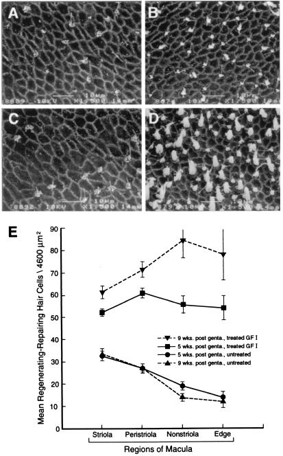
Figure 2.
Treatment with the GF I had the greatest effect on increasing hair cell renewal in the nonstriola and edge areas of the macula. (A–D) Scanning electron micrographs of utricular maculae at 9 weeks postgentamicin exposure. The striola region is represented in A and B—A being ototoxin-damaged, untreated and B receiving 1 month of GF I infusion. The edge region is depicted in C and D—C being a gentamicin-damaged, untreated macula and D a macula receiving 1 month of GF I therapy. A comparison of these micrographs shows that GF I treatment greatly increased the number of renewing vestibular HCs in the edge region. (E) Hair cell counts from the four regions of the macula at 5 and 9 weeks post-gentamicin exposure. [Bars denote ±SEM; 5 weeks (n = 6), 9 weeks (n = 3); scale bars = 10 μm.]

