Figure 4.
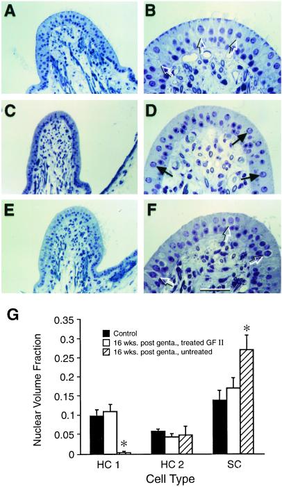
Treatment of gentamicin-damaged inner ears with GF II containing BDNF results in the return of type 1 hair cells in the cristae. (A–F) Photomicrographs of toluidine blue-stained, 1-μm sections of horizontal cristae in the plane of the planum semilunatum at 16 weeks post-gentamicin exposure. (A and B) The normal anatomy of the horizontal crista from a control animal. (C and D) The abnormal anatomy of a gentamicin-damaged horizontal crista that received no additional treatment. (E and F) Normal-appearing anatomy of a horizontal crista from a gentamicin-damaged vestibule after treatment with GF II. Control and ototoxin-damaged GF II-treated cristae show a similar histology with many type 1 HCs present (open arrows) in both A, B, E, and F, whereas the untreated crista in C and D shows a very different histology with few HCs (solid arrows) and no detectable type 1 hair cells. (G) The quantification of cell types in cristae from control; gentamicin-exposed, untreated; and gentamicin-exposed, GF II-treated animals. For each group (n = 3), asterisks = P < 0.01. (Bars denote ±SEM; scale bar = 50 μm in B, D, and F and 100 μm in A, C, and E.)

