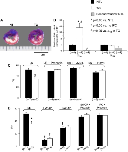
Fig. 3.
Infarct size (IS) and α1A-AR mRNA levels in TGs, NTLs, and NTLs subjected to SWOP (Second window NTL). Symbols indicate statistical differences. The number of the animals (n) is shown in parentheses. Values are means ± SE. A: examples of infarcted hearts from both NTL and TG groups after ischemia-reperfusion (I/R) showing the area at risk (AAR; blue staining) and infarcted tissue [white areas that fail to stain with 2,3,5-triphenyltetrazolium chloride (TTC)]. Note that the infarct was more homogeneous in NTLs and more patchy in TGs, reflecting myocardial salvage in the latter. B: using quantitative real-time reverse transcriptase polymerase chain reaction (qRT-PCR) of cardiomyocyte RNA, compared with untreated NTLs, mRNA expression of the α1A-AR was increased ∼40-fold in TGs but was reduced 10-fold in second window preconditioned NTLs. However, expression of the α1B-AR was similar among NTL, TG, and second window preconditioned NTL groups. C: ratio of IS to AAR (expressed as a percentage and shown on the ordinate as %). In the absence of IPC, IS/AAR in response to I/R was lower in TGs. After administration of the α1-AR blocker prazosin, the NOS inhibitor l-NNA, or the MEK inhibitor U-0126, the cardioprotection from I/R observed in untreated TG rats was abolished, whereas IS/AAR was unchanged in prazosin-treated NTLs. D: IS/AAR after FWOP was reduced in both the NTL and TG groups, but, after SWOP, IS/AAR was only reduced in NTLs. In response to prazosin, given either at the time of preconditioning (IPC + prazosin) or throughout SWOP (SWOP + prazosin), the reduction in IS/AAR observed both in the TG rats and in the NTLs subjected to SWOP was abolished.

