Fig. 1.
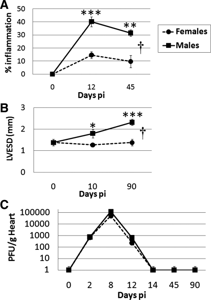
Sex differences in coxsackievirus B3 (CVB3) myocarditis in relation to cardiac function and viral replication. Male and female BALB/c mice were inoculated intraperitoneally with 103 plaque-forming units (PFU) of heart-passaged CVB3 on day 0, and acute [days 10 or 12 postinfection (pi)] and chronic (days 45 or 90 postinfection) myocarditis (percent inflammation) and cardiac function [left ventricular end-systolic dimension (LVESD)] were assessed by histology (A) or echocardiography (B), respectively. C: viral replication in the heart was examined at various time points postinfection using a plaque assay. Student's t-test compared females with males at individual time points: *P < 0.05; **P < 0.01; ***P < 0.001. ANOVA compared females with males over time: †P = 4.12 × 10⁻⁵ in A and †P = 0.008 in B. Data show means ± SE of 7–10 mice/group.

