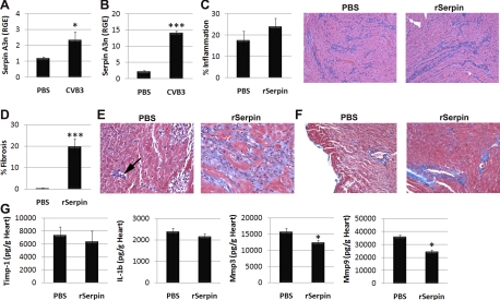
Fig. 4.
Recombinant serpin A 3n (rSerpin) promotes cardiac remodeling during CVB3 myocarditis. Male BALB/c mice received sterile PBS or CVB3 intraperitoneally on day 0, and serpin A 3n levels were examined in the heart by RT-PCR on day 2 (A) or day 10 (B) postinfection. Male BALB/c mice were inoculated intraperitoneally with CVB3 on day 0, and either sterile PBS or rSerpin (10 μg/ml) was injected intraperitoneally every other day from day 1 to day 9 postinfection. Mice were examined at day 10 postinfection. C: summary of histology score (left) and representative hematoxylin and eosin staining for inflammation (purple; right). Magnification: ×64. D: percent fibrosis was calculated as the area with collagen deposition compared with the entire myocardial section using a microscope eyepiece grid. Masson's trichrome to detect collagen deposition (bright blue) showed areas of inflammation (E) or perivascular fibrosis (F). Magnification: ×250 in E and ×100 in F. E: small area of fibrosis (arrow) compared with nonfibrotic inflammation in PBS mice (left) versus widespread fibrosis associated with inflammation in rSerpin-treated mice (right). Data show means ± SE of 10 mice/group in A–C, D, and G. *P < 0.05; ***P < 0.001.

