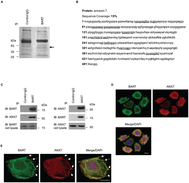
Figure 1. BART binds to ANX7 in lamellipodial-like protrusions.
A. Immunoprecipitates from S2-013 cells using normal rabbit IgG (control) and anti-BART antibody were examined by silver stain analysis. Q-TOF-MS analysis investigated a prominent band in the BART immunoprecipitates (arrow). B. Percent coverage for ANX7 is represented by the identified peptides in the total protein sequence (accession number NM_004034). C. Immunoprecipitated endogenous BART or ANX7 from S2-013 were examined by Western blotting using anti-BART and anti-ANX7 antibodies. Normal rabbit or mouse IgG was used as an isotype control for BART and ANX7, respectively. D. Immunocytochemical staining of S2-013 cells using anti-BART (green) and anti-ANX7 (red) antibodies. Blue, DAPI staining. Bar, 10 µm. E. Arrows indicate that BART (green) and ANX7 (red) colocalize at lamellipodial-like protrusions of S2-013 cells. Blue, DAPI staining. Bar, 10 µm.

