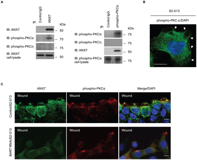
Figure 8. BART supports the colocalization of ANX7 and phospho-PKCα at the leading edges of migrating cells.
A. Immunoprecipitation of ANX7 (left panels) or phospho-PKCα (right panels) from S2-013 cells was examined by Western blotting using antibodies against ANX7, phospho-PKCα and phospho-PKCη. B. Immunocytochemical staining in S2-013 cells, as determined with anti-phospho-PKC antibody (green). Blue, DAPI staining. Arrows, phosphorylated PKC at lamellipodial-like protrusions. Bar, 10 µm. C. Confluent cultures of control and BART RNAi S2-013 cells were wounded. After 4 h, the cells were immunostained using anti-ANX7 (green) and anti-phospho-PKCα (red) antibodies. Blue, DAPI staining. Bars, 10 µm.

