Figure 3.
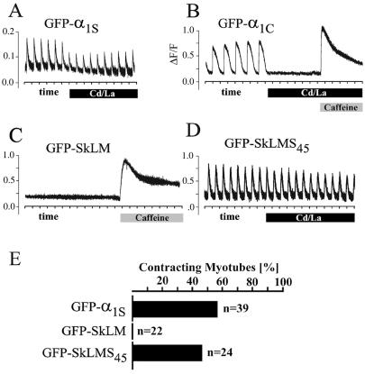
Chimera GFP-SkLMS45 restores skeletal-type EC coupling on expression in dysgenic myotubes. Action-potential-induced Ca2+ transients recorded from dysgenic myotubes expressing DHPR constructs, loaded with the fluorescent Ca2+ indicator Fluo-4 AM. Tick marks on the horizontal axes indicate 2 s. The skeletal GFP-α1S (A) responded to 1-ms stimuli with Ca2+ transients that persisted after blocking currents with 0.5 mM Cd2+ and 0.1 mM La3+ (solid bar), whereas the cardiac GFP-α1C Ca2+ transients (B) were blocked by the Cd2+/La3+ solution. Myotubes expressing GFP-SkLM (C) failed to restore action-potential-induced Ca2+ transients (n = 10 dishes) even though Ca2+ release could be induced with 6 mM caffeine (shaded bar). GFP-SkLMS45 (D) fully restored action-potential-induced Ca2+ transients that were resistant to Cd2+/La3+ block of Ca2+ currents, indicating skeletal-type EC coupling. As for GFP-α1S, the application of Cd2+/La3+ sometimes caused a modest reduction in the amplitude of the transient in cells expressing GFP-SkLMS45. (E) Electrically evoked contractions (100 ms, 100 V) recorded in Cd2+/La3+ from dysgenic myotubes expressing either GFP-α1S, GFP-SkLM, or GFP-SkLMS45 indicated as percentage of myotubes stimulated.

