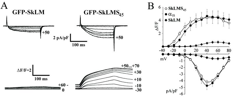
Figure 4.
Restoration of bidirectional coupling by expression of chimera GFP-SkLMS45. (A) Whole-cell Ca2+ currents (Upper) and depolarization-induced Ca2+ transients (Lower) recorded simultaneously from dysgenic myotubes expressing GFP-SkLM or GFP-SkLMS45. Step depolarizations (200-ms pulses) were applied in 10-mV increments from a holding potential of −80 mV after a prepulse protocol (24). The vertical scale indicates ΔF/F, Ca2+-induced Fluo-3 fluorescence increments (ΔF) with respect to basal fluorescence (F). (B) Voltage dependence of depolarization-induced Ca2+ transients (ΔF/F, Upper) and of peak current densities (pA/pF, Lower) recorded from dysgenic myotubes expressing GFP-α1S (●), GFP-SkLM (♦), and GFP-SkLMS45 (○). Values represent the mean ± SEM of 11–20 recordings. The small Ca2+ transients for GFP-SkLM appeared to be a direct consequence of Ca2+ influx through the DHPR (see text).

