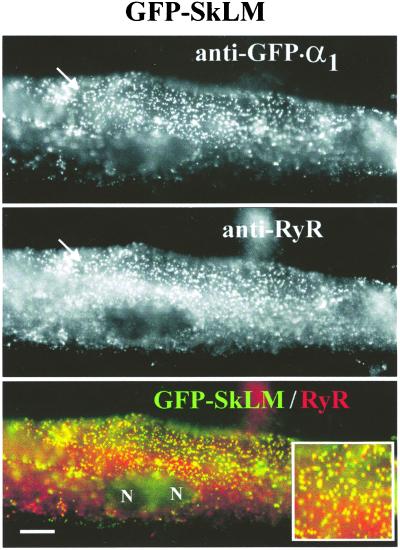
Figure 5.
Reduced Ca2+ currents and lack of EC coupling are not a result of failed junctional targeting of chimera GFP-SkLM. Subcellular localization of chimera GFP-SkLM in a transiently transfected dysgenic myotube (GLT). Double-immunofluorescence labeling was performed with antibodies against GFP, N-terminally fused to the DHPR chimera (Top) and against RyR1 (Middle). The “merged image” (Bottom) emphasizes the colocalization (yellow foci) of GFP-SkLM (green) and RyR1 (red) in clusters that represent junctions of the SR with transverse tubules or with the plasma membrane. Arrows indicate examples of GFP-SkLM/RyR1 colocalization. Inset shows a 2-fold-enlarged view of coclustered channels. N, nuclei. (Bar = 10 μm.)

