Figure 2.
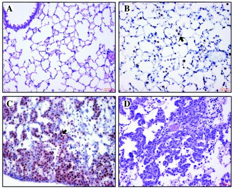
Histological changes in the lungs of GLp65/FGF-3 bitransgenic mice treated with placebo or 500 μg/kg RU486. (A) H&E-stained section of lung from placebo-treated mouse. (B) Immunohistochemical staining for Mac-3 of lung from 1137 bitransgenic mouse treated with RU486 for 6 weeks. Free alveolar macrophages (FAMs) are abundant in the alveoli as indicated by the arrow. (C) Immunohistochemical staining for Nkx2.1 of lung from the 1138H bitransgenic mouse treated with 500 μg/kg RU486 for 2 weeks. Type II epithelial cells are increased strikingly along the septa and in alveoli as indicated by the arrow. (D) H&E section of lung from the 1292 bitransgenic mouse treated with 500 μg/kg RU486 for 1 week.

