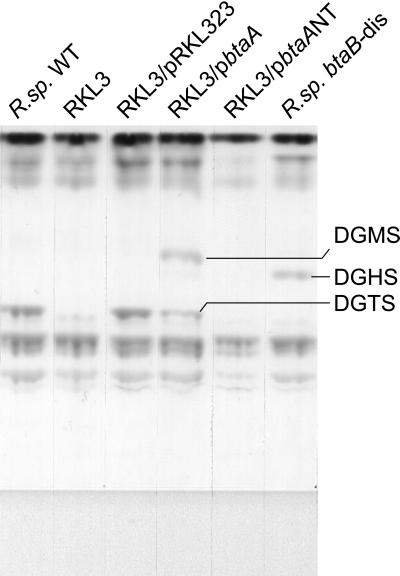
Figure 2.
Comparison of lipid extracts from different strains of R. sphaeroides. All cells were grown under phosphate-limited conditions at an initial Pi concentration of 0.1 mM. A one-dimensional thin-layer chromatogram stained by iodine vapor is shown. The indicated strains and plasmids are described in Table 1 and in the text.

