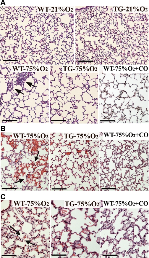
Fig. 5.
Lung histological examination showing the effects of hyperoxia, HO-1 overexpression and CO inhalation on alveolarization, hemorrhage, and edema. A: paraffin lung sections stained with hematoxylin and eosin demonstrated increased alveolar size and reduced septation in WT, HO-1 TG, and CO-treated mice exposed to hyperoxia (75%O2) 2 wk after birth, compared with normoxic (21% O2) mice. WT lungs also exhibited abundant perivascular mononuclear infiltration (arrows). B: areas of hemorrhage (seen as mild to moderate accumulation of red blood cells in the pulmonary parenchyma) as well as thickened alveolar walls, indicative of pulmonary edema (C, arrows) were abundant in hematoxylin and eosin-stained lung sections from WT mice exposed to 75% O2 but less common in lung sections from similarly exposed HO-1 TG and CO-treated mice (n = 4–6 mice per group). Scale bars in A and B = 100 μm and in C = 50 μm.

