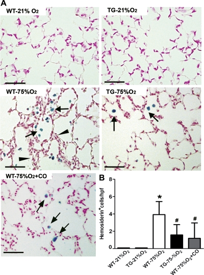
Fig. 7.
Hemosiderosis induced by hyperoxia exposure is attenuated by HO-1 overexpression and CO inhalation. A: lung sections from WT, TG, and CO-treated mice stained with Perls Prussian blue iron revealed areas of severe hemosiderosis (arrows) in proximity to thickened alveolar walls (arrowheads) in WT mice exposed to 75% O2. Only moderate hemosiderosis was observed in hyperoxic HO-1 transgenic and CO-treated mice. B: hemosiderosis was quantified by counting iron-laden cells per high-power field in lung sections from each experimental group (n = 8). *P < 0.05 vs. WT- 21% O2; #P < 0.05 vs. WT-75% O2. Scale bar = 50 μm.

