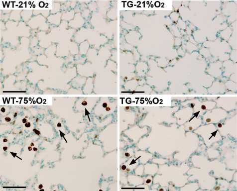
Fig. 9.
Hyperoxia-induced HO-1 expression is mainly restricted to pulmonary alveolar macrophages. Immunohistochemistry of paraffin lung sections with an antibody against murine HO-1 demonstrated minimal staining in mice that were exposed to room air (21% O2), whereas immunostaining was intense in macrophages (arrows) of both genotypes exposed to hyperoxia (75% O2) with significantly fewer stained macrophages in the hyperoxic HO-1 TG lungs, compared with WT hyperoxia. The evident epithelial HO-1 signal in the TG lung was due to cross-reactivity with the human HO-1 transgene expressed in type II epithelial cells. Scale bar = 50 μm.

