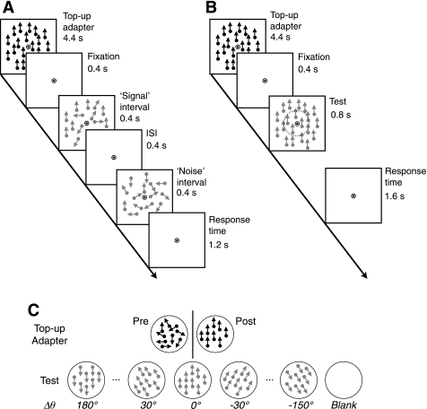
Fig. 1.
Stimuli and procedures for psychophysical and functional magnetic resonance imaging (fMRI) experiments. A: schematic diagrams of visual stimuli presented during psychophysical experiments. Each 7.2-s trial started with a high-contrast top-up adapter, which was followed by the “signal” and “noise” intervals in a random order. Subjects indicated which interval contained signal dots within 1.2 s after the offset of the second interval. ISI, interstimulus interval. B: schematic diagrams of visual stimuli during fMRI experiments. The adapter was identical to that in the psychophysical experiments. Subjects indicated whether or not motion directions were the same between the central and peripheral annulus. The imaginary circle with a broken line indicates the boundary between the 2 annuli. C: top-up adapter and test stimuli during fMRI experiments. High-contrast dots in a top-up adapter were moving in random directions before adaptation and in a single direction after adaptation. Low-contrast dots in the test period were moving always in a single direction. The relative direction difference between the adapter and test stimulus (Δθ) was varied at the 7 levels ranging from 0° to 180°. In fMRI experiments, “blank” trials were inserted randomly in between regular trials.

