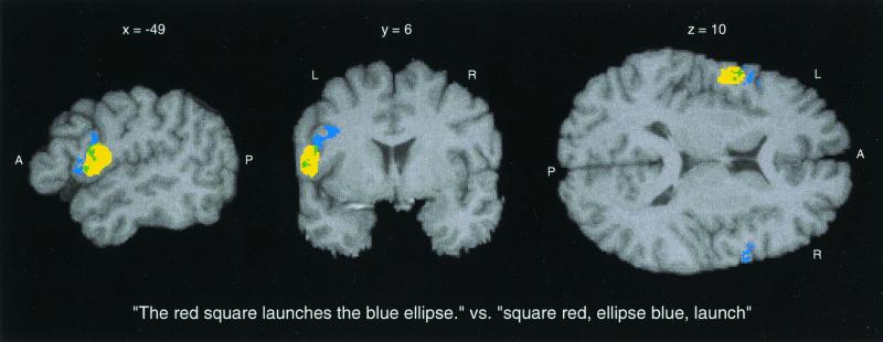
Figure 3.
Cortical activation of sentence relative to single word utterances. Significantly activated voxels are projected in yellow onto anatomical MR sections of a reference brain. For anatomical comparison, voxels belonging to BA 44 are projected in blue on the same reference brain. A smaller anterior portion of the activated volume (16.3%, shown in green) overlaps BA 44, and the larger part of the activation (83.7%) lies caudally adjacent to BA 44, most probably corresponding to the Rolandic part of BA 6. The maximally activated voxel was located at x = −54, y = 6, z = 10 (coordinates as given by spm96). (Note that the depicted sagittal section is taken more medially to improve the visibility of the anatomical configurations of the posterior inferior frontal gyrus).

