Fig. 2.
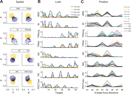
Variation of elbow and shoulder muscle activation across target force directions evaluated in the spatial, load, and position protocols (A–C, respectively) for subject S4. A: spatial tuning of EMGs underlying generation of force in 210 target directions (each circle represents data from a single trial). B: EMGs recorded during the load protocol scaled with load level, whereas spatial tuning was relatively unaffected. MLF, maximum lateral force. C: relative levels of muscle activation varied depending on the force direction (shown along the x-axis) and the position of the hand (indicated in the legend). Force directions and hand positions for C are identified in Table 1 and Fig. 1, A and B, respectively. Muscle names are indicated in an abbreviated form: BRD, brachioradialis; BI, biceps brachii; TRIlong and TRIlat, long and lateral heads of triceps brachii, respectively; AD, MD, and PD, anterior, medial, and posterior fibers of deltoid, respectively; and PECTclav, clavicular fibers of pectoralis major.

