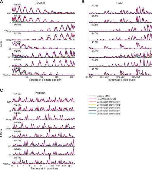
Fig. 9.
Reconstruction of EMGs for the spatial, load, and position protocols (A, B, and C, respectively) by linear combinations of 4 or 5 synergies based on data for subject S4. The original EMGs (dashed black lines), reconstructed data (solid magenta lines), and contribution of each muscle synergy (common color scheme used for associated synergies) to the reconstruction are shown. The associated synergies are displayed in the 1st, 2nd, and 7th columns of Fig. 5. The muscle VAF value is indicated for each associated muscle. B and C: the 54 and 8 trials at each load level and hand position are shown, respectively. All trials are ordered along the x-axis in the trial sequence shown in Figs. 1D and 2, B and C.

