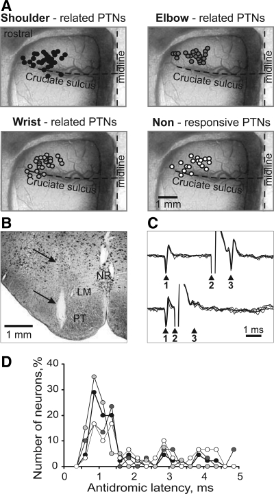
Fig. 2.
Location of PTNs and their identification. A: area of recording in the forelimb representation of the left motor cortex. Microelectrode entry points into the cortex are combined from all cats and shown by circles on the photograph of cat 9 cortex. Tracks where PTNs with shoulder-related, elbow-related, and wrist-related receptive fields and nonresponsive PTNs were recorded are shown by black, dark gray, light gray, and white circles, respectively. B: reference electrolytic lesion in the left pyramidal tract made with the stimulation electrode in cat 8. Gliosis surrounding the electrode track and the reference lesion are indicated by arrows. The electrode was positioned approximately at the Horsley-Clarke rostro-caudal coordinate of P7.5. LM, lemniscus medialis; NR, nucleus raphes; PT, pyramidal tract. Frontal 50-μm-thick section, cresyl violet stain. C: collision test determines whether PTN response is antidromic. Top: the PTN spontaneously discharges (arrowhead 1), and the pyramidal tract is stimulated 3 ms later (arrowhead 2). The PTN responds with latency of 1 ms (arrowhead 3). Bottom: the PTN spontaneously discharges (arrowhead 1), and the pyramidal tract is stimulated 0.7 ms later (arrowhead 2). The PTN does not respond (arrowhead 3) because in 0.7 ms its spontaneous spike was still en route to the site of stimulation in the pyramidal tract, and thus collision/nullification of spontaneous and evoked spikes occurred. D: distribution of latencies of antidromic responses to stimulation of the pyramidal tract of PTNs of different groups. Shoulder-related, elbow-related, wrist-related, and nonresponsive PTNs are denoted by black, dark gray, light gray, and white circles, respectively.

