Figure 9.
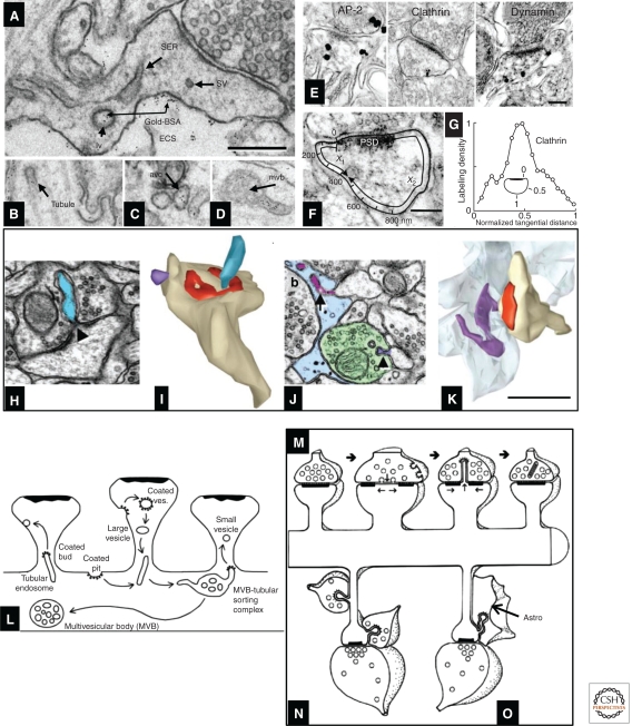
Endocytosis and trans-endocytosis at dendritic spines. (A) SER, small vesicles (sv), and recycling endosomes; the latter may be identified by their content of gold-BSA endocytosed from the extracellular space (ECS) into large vesicles (lv), (B) tubules, (C) amorphous vesicular clumps, and (D) multivesicular bodies. (E–G) Silver-enhanced pre-embedding immunogold identifies three proteins (AP-2, clathrin, and dynamin) critical for endocytosis; label for all three concentrates close to the spine plasma membrane. To assess their position with respect to the synapse, the locus of each particle was projected onto the spine plasma membrane as illustrated in F, allowing computation of a normalized tangential distance along the plasma membrane. As shown for clathrin in G, each of these endocytic proteins concentrated in a sector of the spine lying roughly halfway between the edge of the PSD and the point on the spine plasma membrane closest to the spine neck opposite to the PSD (illustrated as “1” in the inset) (for further description, see Racz et al. 2004). (H) Spinule (blue) emerging from the middle of a perforated PSD (triangle) on a dendritic spine head; this spinule is engulfed by the plasma membrane of the presynaptic axon. (I) 3D reconstruction of the spinule in H (blue), and also a second smaller spinule (purple) being engulfed by a neighboring axon, not shown). (J) Spinule from a dendritic spine (purple arrow, top) being engulfed by a perisynaptic astroglial process (light blue), and a second smaller spinule from a different spine (purple arrowhead, bottom) being engulfed by a neighboring axon (green), similar to the purple reconstruction in I. (K) 3D reconstruction of the astroglial-engulfed spinule shown on a single section in J (spinule [purple], astroglial process [light blue], spine [tan], PSD [red]). (L–O) Contrasting endocytosis and trans-endocytosis. (L) A single MVB-tubular sorting complex in the dendritic shaft serves multiple dendritic spines, where coated pits and vesicles are endocytosed from some spines, and small vesicles bud off (also via coats) from tubular endosomes, to be exocytosed at the plasma membrane of neighboring spines. (M) Trans-endocytosis from a spine to the presynaptic axon may serve to transmit signaling molecules or to remove perforations following sustained presynaptic activation. (N) Trans-endocytosis by neighboring axons may reflect competition for the postsynaptic spine. (O) The function of trans-endocytosis by neighboring astroglial processes is unknown. Scales, (A–D, H–K) 500 nm; (E, F) 200 nm. (Panels A–D and L are from Cooney et al. 2002; reprinted, with permission, from the authors; panels E–G are from Racz et al. 2004; reprinted, with permission, from the authors; and panels H–K and M–O are from Spacek and Harris 2004; reprinted, with permission, from the authors.)

