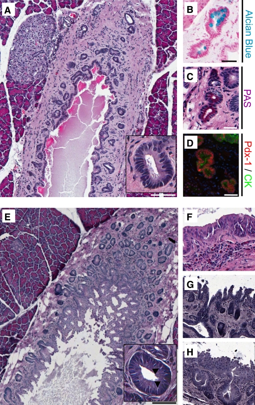
FIG. 1.
The extent and frequency of PDGs surrounding the main pancreatic duct are increased by exendin-4 treatment in rats. Sections from the head of the pancreas from an untreated control rat (A) and after 12 weeks of daily exendin-4 injections (E), in which PDG clusters were identified surrounding the main pancreatic duct. PDGs were confined to the mesenchyme surrounding the main duct in controls but, after exendin-4, expanded to the extent that they projected into the lumen of the pancreatic duct as complex villous-like structures. A and E, insets: PDG cells were columnar in comparison with the cuboidal ductal cells and included goblet-like cells (arrowheads). B and C: PDGs contained mucin confirmed by Alcian blue and PAS staining. D: In contrast to duct cells, PDG cells also expressed Pdx-1 (red; combined staining with the duct cell marker cytokeratin [CK] in green). E: PDGs were more common in exendin-4–treated rats (Table 1). F–H: In addition, the epithelium often showed pseudostratification and pseudopapillary features, which are features characteristic for PanIN-like lesions. Scale bars = 200 μm (A and E) and 100 μm (B–D), and magnification ×20 (F–H). (A high-quality digital representation of this figure is available in the online issue.)

