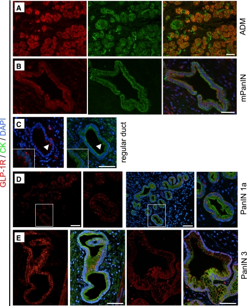
FIG. 6.
GLP-1R expression is present in PanIN lesions in Pdx1-Kras mice and humans. GLP-1R (red; shown with combined cytokeratin [CK] labeling in green) was detected in areas of acinar-to-ductal metaplasia (ADM) (A) and mPanIN lesion (B) in the pancreas of Pdx1-Kras mice. Colocalization of GLP-1R and cytokeratin is indicated in the merged images by the color orange. C: In human pancreas, GLP-1R expression was more apparent in the columnar cells (arrowheads) in regular ducts compared with adjacent normal cuboidal duct cells shown away from the arrowhead. D: Where duct cells adopt the columnar phenotype (PanIN1a lesion shown), GLP-1R expression becomes more apparent. E: In more advanced PanIN3 lesions, GLP-1R immunoreactivity also was clearly present. Scale bars = 100 μm. (A high-quality digital representation of this figure is available in the online issue.)

