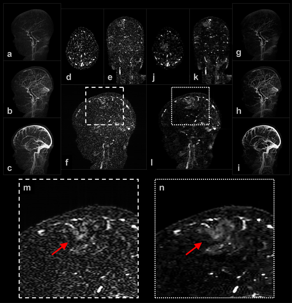
Figure 3.
Selected images from Example 1: (a–c) are sagittal MIPs of early, middle, and late filling stage volumes from a view-shared SENSE+Homodyne reconstruction of the neurovasculature of a pediatric patient, and (d–f) are axial, coronal, and sagittal slices from a late-filling stage SENSE+Homodyne volume; (g–i) and (j–l) are the corresponding images from the NCCS reconstructions. (m) and (n) are the respective enlargements of a region of interest (ROI) in (f) and (l).

