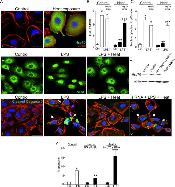
Figure 1. Hsp70 induction limits TLR4 signaling in enterocytes.
A: Confocal micrographs showing the expression of Hsp70 (green), β-actin (red) and DAPI (blue) in IEC-6 enterocytes that were either untreated (i) or exposed to 42°C for 45 minutes (ii). B–C: quantitative RT-PCR showing the expression of IL-6 (B) or quantification of the extent of NFkB translocation (C) in IEC-6 cells that were either untreated (white bars) or exposed to heat (black bars), and were either untransfected or were transfected with Hsp70 siRNA. *p<0.05 vs untreated control; **p<0.05 vs heat-control, ***p<0.001 vs control cells transfected with Hsp70 siRNA. D: Confocal micrographs of IEC-6 enterocytes that were either untreated (i), treated with LPS (50μg/ml, 45 min, ii) or were treated with LPS after pre-treatment with heat (iii). Quantification in C is based upon over 50 cells per field and over 50 fields examined in 4 separate experiments. *p<0.05 vs untreated control; **p<0.05 vs heat-control. E: Representative SDS-PAGE showing lysates of IEC-6 that were untreated (control), incubated with PBS alone (vehicle), or were transfected with either control siRNA against no known substrate (non-targeted siRNA) or siRNA to Hsp70 (Hsp70 siRNA). Blot was stripped then re-probed with antibodies to β-actin. F: confocal micrographs (i–iv) and quantification (v) of IEC-6 enterocytes that were either untreated (i), treated with LPS in the absence (ii) or presence of pre-exposure to heat as above (iii), or pre-treated 48 hours prior with siRNA to Hsp70 as in (iv). *p<0.05 vs control; **p<0.005 vs LPS control; ***p<0.05 vs heat+hsp70sirna saline. Representative images are taken of over 50 fields examined with over 50 cells/field in 4 separate experiments. Size bar = 10μ. Representative apoptotic cells are indicated by arrows.

