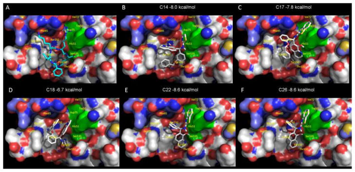
Figure 5. Molecular docking of compounds 14 and 26 onto WNV protease structure.
The WNV protease crystal structure coordinates (PDB ID code 2FP7) were used for molecular docking. AutoDock Vina was used to dock each compound in the active site bound by Bz-Nle-Lys-Arg-Arg-H substrate-based inhibitor (Erbel et al., 2006). The solvent-accessible surface of the protein is shown in color-coded atoms (white, carbon; red, oxygen; blue, nitrogen). The catalytic triad (H51, D75, and S135) is shown as a green surface. A. The structure of the Bz-Nle-Lys-Arg-Arg-H substrate-based inhibitor bound WNV NS2B-NS3pro (Erbel et al., 2006). B. Compound 14 is shown in the orientation posed by molecular docking. Compounds 17, 18, 22 and 26 are shown in panels C, D, E and F, respectively.

