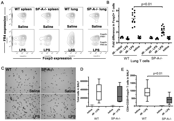
Figure 1. SP-A enhances the formation of Foxp3+Tregs in the lung on extended LPS stimulation in vivo.
LPS O55:B5 was instilled oro-pharyngeallyonce, for a duration of 0, 2 or 8 days, and levels of Foxp3+, FR4-hi and FR4-intermediate cells in lungs or spleen of these mice were detected by flow cytometry(A). The fold induction in Foxp3+ T cells in individual mice (data points) are obtained after normalization to the unchanged proportions of Foxp3+ T cells in spleens of corresponding mice across5 independent experiments (B). C and D, Cytospins and differential T cell counts of BALF obtained from total lung volume lavage of saline or LPS treated mice after 8 days of exposure. Absolute numbers of CD4+CD25+Foxp3+ Tcells present in BALF collected from total lung volume lavage were also determined by flow cytometric analysis (E). The box-and-whiskers plots (D and E) show both the range of cell numbers from highest to lowest, as well as lower quartile, median and upper quartile.

