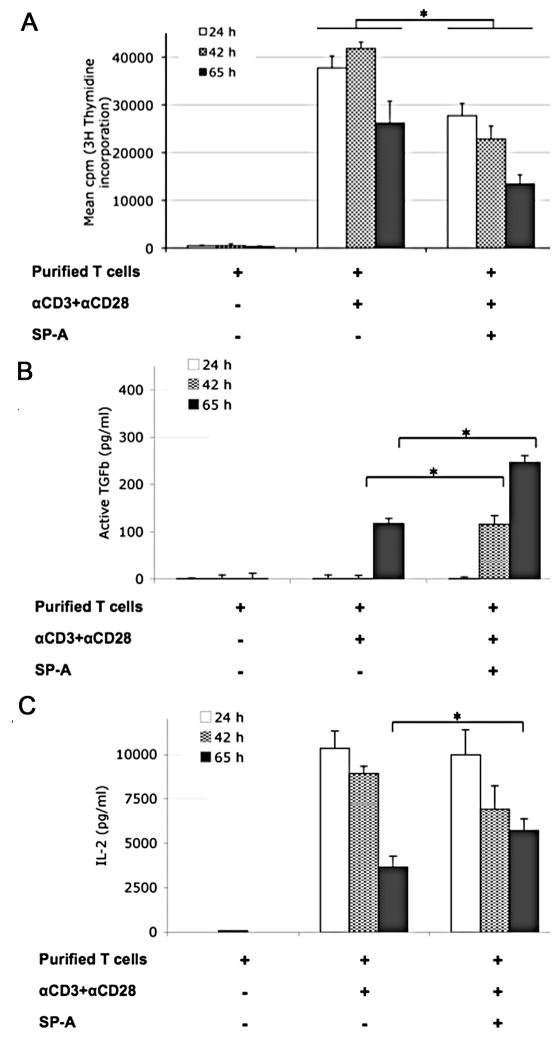
Figure 3. SP-A enhances IL-2 and bio-active TGFβ production in extended culture, even as T cell proliferation is suppressed in a kinetic manner during initial activation.
Purified mouse T cells were left unstimulated or stimulated for the indicated times with anti-CD3 and anti-CD28 in the absence or presence of SP-A (20 μg/ml). (A) Mean cpm of [3H]-thymidine incorporation over 15 h at the indicated time points, by proliferating cells from replicate wells in a representative experiment is depicted. (B) TGFβ levels from culture supernatants were determined by a functional bioassay using CCL64MLE cells as described in Methods.(C) IL-2 levels in culture supernatants at the indicated time points were measured by ELISA. * p<0.05 for fold differences across corresponding time points in three independent experiments by the Mann-Whitney test.

