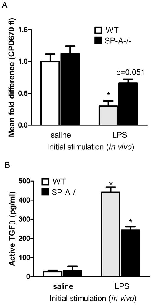
Figure 5. SP-A enhances functional Foxp3+Tregs on extended LPS stimulation in vivo.
LPS was instilled oro-pharyngeally for 8 days, and T cells were harvested and purified from lungs. These cells were used 1:1 in a suppression assay with freshly isolated T cells pulsed with CPD670, and the fold difference in proliferating cells (≥2 divisions) in relation to labeled, activated target cells was calculated (A). A lower level of fluorescence is indicative of greater extent of proliferation, and lower degree of suppression. One lobe of the lung from saline or LPS treated mice was used to prepare lysates for determining amounts of total and active TGFβ levels by ELISA (B). *p<0.05 compared to respective saline treatment condition.

