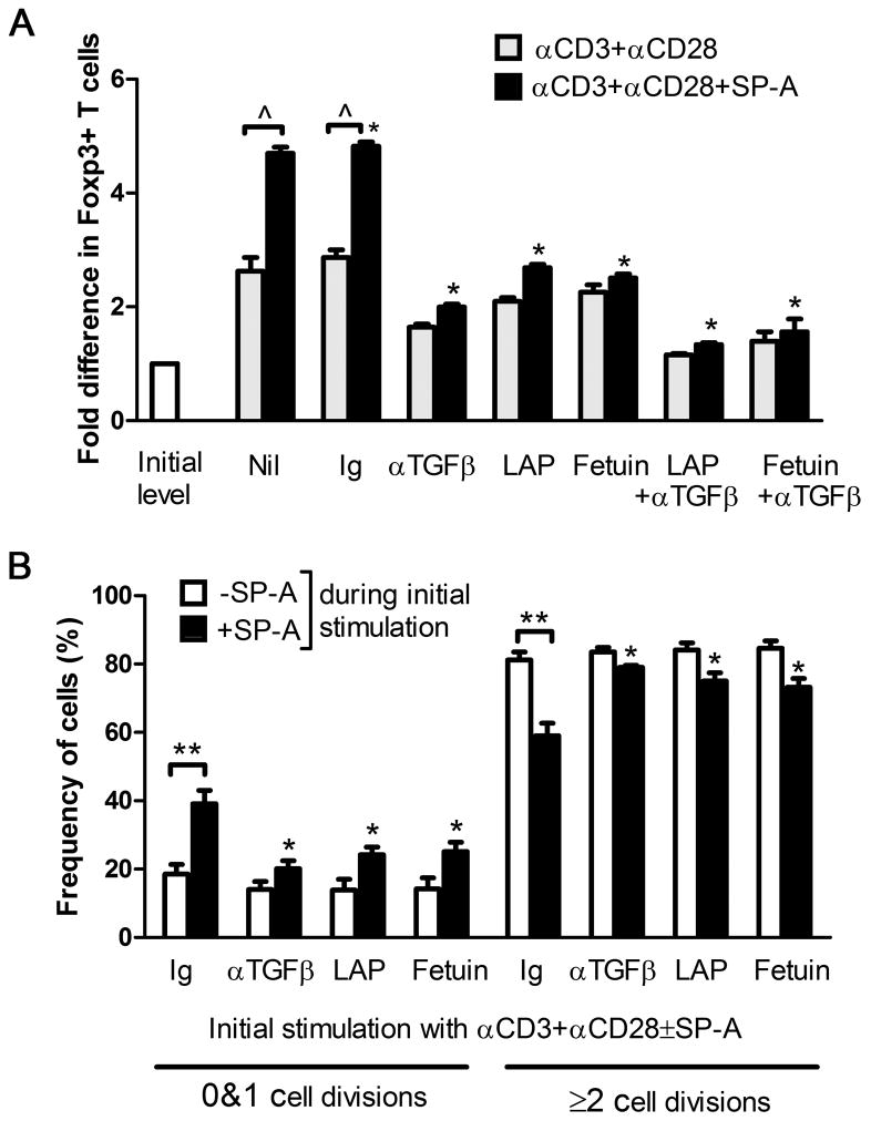
Figure 6. TGFβ blockade abrogates SP-A induced Foxp3+Tregs and restores T cell proliferation in an ex vivo suppression assay.
Foxp3 levels were detected by flow cytometry after 70 h of culture in the absence (gray) or presence (black) of SP-A, with control Ig or TGFβ blocking agents as indicated (A). Data is depicted as fold difference in Foxp3+ cells compared to mean initial levels from pooled untreated mice. *p<0.05 with respect to the activated, control Ig+SP-A group across treatment conditions, ^ p<0.01 in −/+SP-A treatment. Suppression assays were performed with anti-CD3+anti-CD28 stimulation using cells subject to initial stimulation in the absence or presence of SP-A (B). The proportion of cells that have either remained undivided or have undergone 2 or more divisions in the suppression assay following initial stimulation under indicated treatment conditions is depicted. *p<0.05 with respect to control Ig+SP-A across treatment conditions, ** p<0.02 in −/+SP-A treatment.

