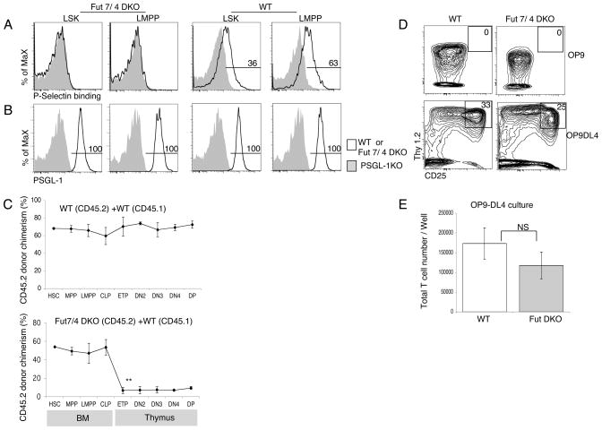
Figure 6.
Fucosyltransferase enzyme play role in thymus settling process. (A) BM from Fuc-TVII/Fuc-TIV DKO (left panels) and wild-type (WT) (right panels) mice was analyzed by flow cytometry for Functional PSGL-1. (B) BM from Fuc-TVII/Fuc-TIV DKO (left panels) and wild-type (WT) (right panels) mice was analyzed by flow cytometry for surface PSGL-1 protein expression using mAb to PSGL-1. Numbers represent the percentage of WT cells in the indicated gate. (C) Mixed BM chimeras were generated using CD45.2 WT BM and CD45.1 WT BM mixtures as controls (top panel), CD45.2 Fuc-TVII/Fuc-TIV DKO BM and CD45.1 WT BM mixtures (bottom panel). Chimeras were analyzed by flow cytometry after 10 weeks using antibodies to CD45.1 and CD45.2 to determine chimerism. The mean CD45.2 donor chimerism ± S.E.M. for each indicated population is shown. (D) Sorted LMPP from WT or Fuc-TVII/Fuc-TIV DKO mice was cultured on OP9 (top panel) or OP9-DL4 (bottom panel) stromal layers in triplicate. After 10 days, cultured cells were analyzed by flow cytometry for expression of CD25 and Thy1.2. Representative FACS plots of cells gated on CD45+ are shown. (E) Total numbers of cells obtained from the cultures described in panel D for OP9-DL4. Data are mean and ± S.E.M. of three wells.

