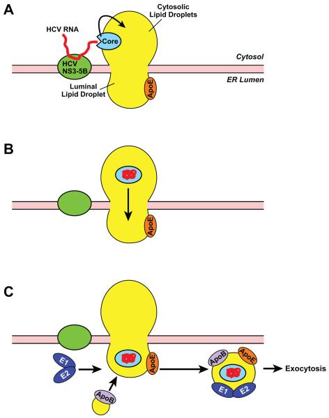
Figure 2.
A hypothetic model illustrating HCV assembly. HCV RNA is synthesized by HCV replication complex composed of viral proteins NS3-NS5B at the cytosolic face of the ER membranes. The viral RNA then binds to the core protein localized at the surface of the lipid droplets adjacent to the viral replication complex (A). Upon binding with viral RNA, core goes through a conformational change so that the core-RNA complex enters the core of cytosolic lipid droplets, allowing the complex to reach lipid droplets in the ER lumen by traveling through ER domains enriched in neutral lipids (B). The HCV-containing luminal lipid droplets fuse with apoB, acquire E1 and E2 heterodimer, and are secreted out of cells through exocytosis (C).

