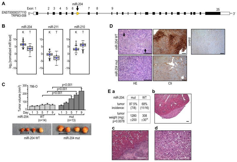
Figure 1. MiR-204 has tumor-suppressing activity in renal clear cell carcinoma.
(A) Localization of miR-204 at the 5′ end of intron 6 of the host gene, TRPM3.
(B) Expression of miR-204, its paralog miR-211, and miR-210 in human ccRCC as compared with normal kidney tissue. Quantification of the indicated normalized miR levels (log2) of the total unpaired kidney (K; n=114) and tumor (T; n=128) samples are shown. The boxes represent lower and upper quartiles separated by the median (thick horizontal line) and the whiskers extend up to the minimum and maximum values, excluding points that are outside the 1.5 interquartile range from the box (marked as circles). Means ± SD of each distribution are indicated by closed dots and crosses on the whiskers, respectively. All differences were statistically significant at p<0.001.
(C) Growth of pre-formed 786-O VHL(−) tumors in subcutaneous xenografts injected every other day over a period of 9 days with lentiviral particles of wild-type pre-miR-204 (WT) or pre-miR-204 with the 3-base-pair mutation (mut). The data are cumulative over two independent series of injections. Representative samples from each group of tumors are shown below. Scale bar = 1 cm.
(D) Representative images of H&E-stained or C9 antibody-probed sections from tumors injected with wild-type (top) or mutant (bottom) pre-miR-204. A section from a non-injected tumor stained for C9 is shown as a control. Scale bars = 200 μm.
(E) Incidence and size of RCC tumors resulting from transduced cells injected under the kidney capsules of nude mice (a). Example of H&E-stained sections of tumors formed by cells transduced with wild-type pre-miR-204 (b and c) or with mutant pre-miR-204 (d). Scale bar = 200 μm. See also Figure S1.

