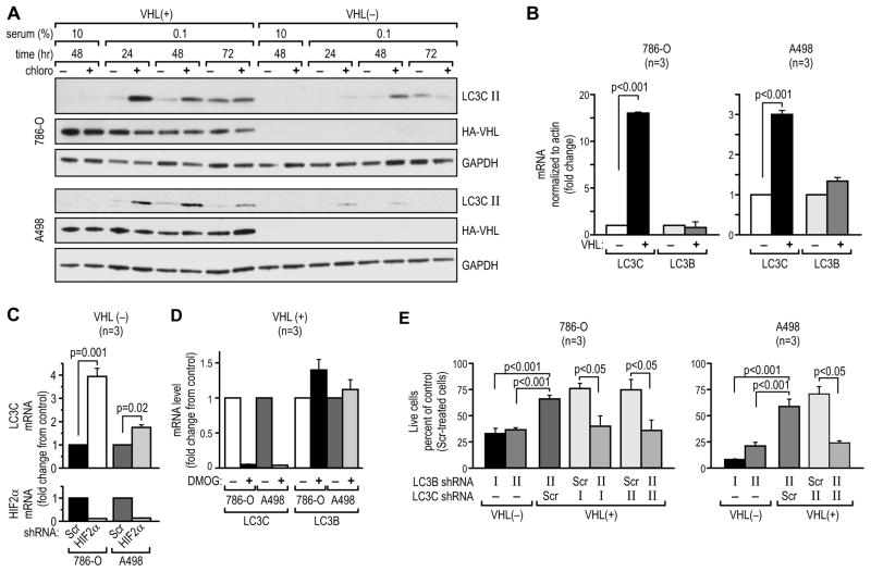
Figure 7. VHL-induced expression of LC3C protects RCC cells from inhibition of LC3B-dependent autophagy.
(A) Western blot showing time course of LC3C-II induction in VHL(+) and VHL(−) 786-O and A498 cells in response to serum starvation.
(B) Quantitative RT-PCR for LC3C and LC3B mRNAs (48 hr of starvation) in VHL(+) as compared with VHL(−) cells.
(C) Quantitative RT-PCR for LC3C mRNA in VHL(−) cells where HIF-2α was knocked down by using siRNA.
(D) Quantitative RT-PCR for LC3C mRNA in VHL(+) cells where HIF was stimulated with DMOG (1 mM) for 16 hr.
(E) Effect of single LC3C or LC3B knockdown or of double LC3C/LC3B knockdown on the viability of indicated cells, as compared with scramble-treated cells. The timeline followed was the same as that shown in Figure 3A. See also Figure S7.

