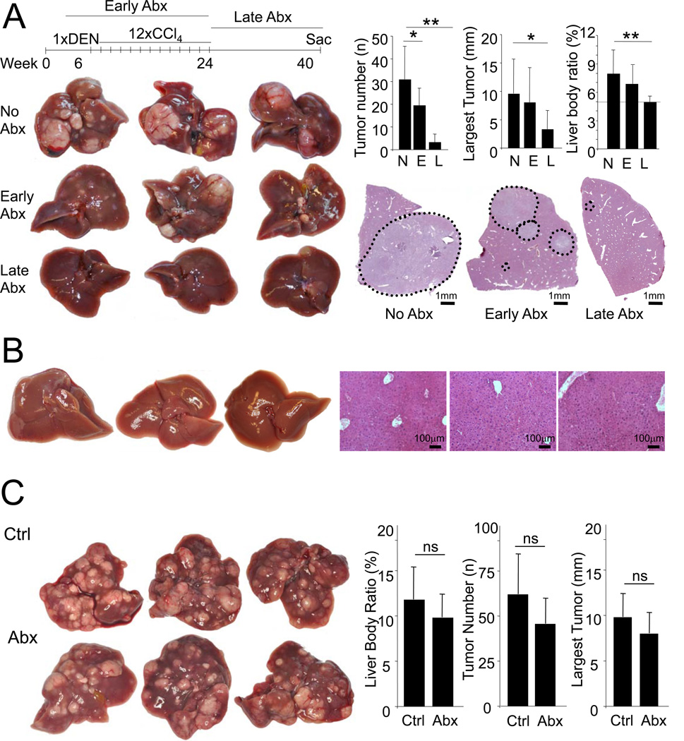
Figure 7. The intestinal microbiota promotes HCC in late stages of hepatocarcinogenesis.
A. WT mice (C3H/HeOuJ) were treated with DEN (100 mg/kg i.p. at the age of 6 weeks) and 12 CCl4 (0.5 ml/kg i.p.) injections and were either not gut-sterilized (“N”, n=7), gut-sterilized starting 2 weeks before DEN injection until 2 weeks after the last CCl4 injection (“E”, n=10), or 2 weeks after the last CCl4 injection until they were sacrificed (“L”, n=8) followed by determination of tumor number, size and liver-body weight ratio. B. WT mice treated as described above were sacrificed at the time point when the gut-sterilization treatment was started to determine whether macroscopic or microscopic tumors were present. C. WT mice were treated with DEN (25 mg/kg i.p.) at day 15 postpartum followed by 14 weekly injections of CCl4. After confirming the presence of tumor by ultrasound (data not shown), mice were treated with (“Abx”, n=7) or without (“Ctrl”, n=6) antibiotics (ampicillin, vancomycin, neomycin and metronidazole) starting 2 weeks after the last CCl4 injection. After 6 weeks of antibiotics, mice were sacrificed and tumor number, size and liver-body weight ratio was determined. Data are represented as means ± SD. * p<0.05, ** p<0.01. ns, non-significant. See also Figure S5.

