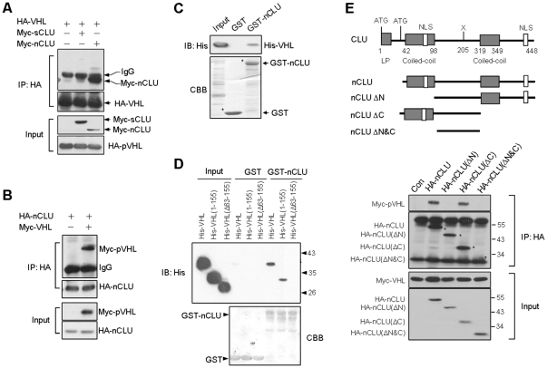
Figure 1. pVHL binds to nCLU.
(A) 293T cells were transfected as indicated. In 24 h, the cells were harvested and cellular proteins were prepared. Immunoprecipitation was performed using HA antibody. (B) 293T cells were transfected with Myc-VHL and HA-nCLU. In 24 h, the cells were harvested and immunoprecipitation was performed using HA antibody. (C) Direct interaction between nCLU and pVHL. Equal amount of bacterial lysates containing His-pVHL were incubated with the glutathione-sepharose beads that already captured GST or GST-nCLU. The beads were washed and His-pVHL retained on beads was determined by immunoblotting. CBB, Coomassie Blue Staining. (D) nCLU bound to pVHL at β domain. Equal amount of bacterial lysates containing His-pVHL, His-pVHL(1-155) or His-pVHL(Δ63-155) were incubated with the glutathione-sepharose beads that already captured GST or GST-nCLU. The beads were washed and His-pVHL or the truncated His-pVHL retained on beads was determined by immunoblotting. (E) A various constructs encoding different nCLUs were designed. 293T cells were transfected with Myc-VHL and nCLU construct as indicated. 24 h post-transfection, the cells were harvested for immunoprecipitation using HA antibody.

