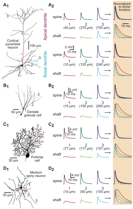
Figure 4. Spines standardize EPSP properties in morphologically realistic neurons.
A1–D1) Morphology of reconstructed neurons: A1) layer 5 pyramidal neuron from the medial prefrontal cortex, B1) hippocampal dentate granule cell, C1) cerebellar Purkinje neuron, and D1) striatal medium spiny neuron. Synaptic inputs were placed onto shafts and spines of the colored dendrites at proximal, intermediate, and distal locations as indicated by the numbered locations (1 to 3). A2–D2) Left, local EPSPs recorded in spines (top traces) or in dendritic shafts (lower traces) at the locations indicated in the different morphologies. Normalized and superimposed traces, expanded in time and shaded at far right, allow comparison of EPSP kinetics. The time course of the underlying synaptic conductance is indicated by dashed lines.

