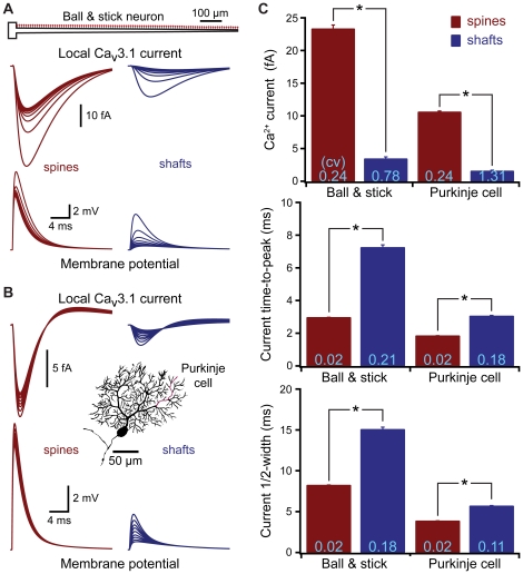
Figure 5. Spines standardize synaptic activation of voltage-gated calcium channels.
A) Top, ball and stick model neuron. Local EPSPs (bottom) and calcium currents (middle) generated by inputs onto spines (left) or dendritic shafts (right) located at ∼100 µm intervals along the dendrite, each synapse contains the equivalent of ten Cav3.1 (T-type) calcium channels (total maximum conductance, 50 pS). B) Local EPSPs (bottom) and calcium currents (top) generated by inputs onto spines (left) and shafts (right) located at ∼10 µm intervals along a spiny dendrite (red) of a cerebellar Purkinje neuron (inset). C) Average calcium current amplitude, time-to-peak, and half-width for all spine (red) and shaft (blue) inputs in the ball and stick (n = 100 inputs) and Purkinje neuron (n = 367 inputs) models. Asterisks indicate p<0.0001 (paired t-tests). CVs are indicated in light blue at base of each bar.

