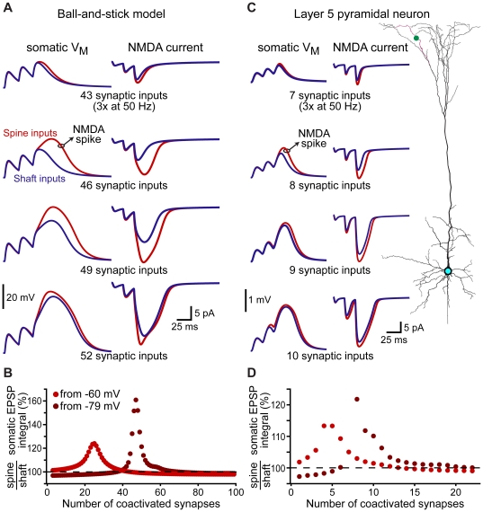
Figure 7. Spines lower the threshold for NMDA spike generation.
A) Somatic voltage (left) and NMDA current (right) evoked by simultaneous activation of different numbers of AMPA+NMDA inputs (trains of 3 activations at 50 Hz) at synaptic locations evenly distributed along the dendrite of a ball-and-stick model resting at −79 mV. For each trial, NMDA currents were recorded from the synaptic input closest to the half-way point along the dendrite. Blue traces reflect responses to shaft inputs, while red traces are responses to spine inputs. B) Plot of the ratios of somatic EPSP integrals (spine inputs/shaft inputs) for trains of different numbers of evenly distributed inputs when the resting potential was set to −79 mV (brown) or −60 mV (red). C) Somatic voltage (left) and NMDA currents (right) evoked by trains of different numbers of AMPA+NMDA synaptic inputs (3 activations at 50 Hz) evenly distributed along the indicated apical branch of a layer 5 pyramidal neuron (right; red dendrite, green dot placed at half-way point along branch). D) Summary plot of the ratios of somatic EPSP integrals (spine inputs/shaft inputs) for trains of different numbers of coactivated inputs in the layer 5 pyramidal neuron resting at −79 mV (brown) or −60 mV (red).

