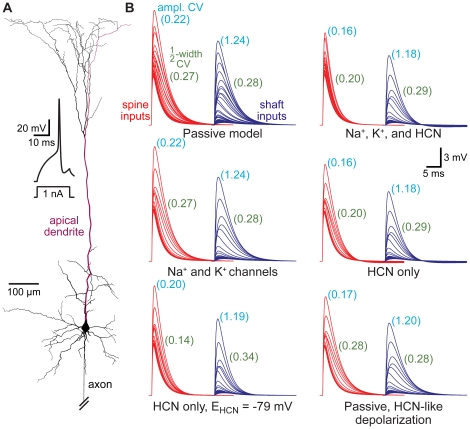
Figure 9. Dendritic HCN channels enhance spine-dependent standardization of EPSPs.
A) Reconstructed layer 5 pyramidal neuron from the somatosensory cortex with spines at ∼10 µm intervals throughout the dendritic tree. Inset, action potential generated in an “active model" containing sodium, potassium, and HCN channels. B) EPSPs generated in spines (red) or shafts (blue) at ∼50 µm intervals along the apical dendrite (red dendrite in A) in models with different passive and active properties. Numbers in light blue and green indicate coefficients of variation (CVs) for EPSP amplitudes and half-widths, respectively, for all local responses (10 µm intervals) to spine and shaft inputs in the various models.

