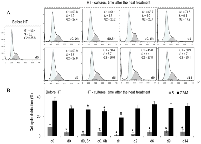
Figure 5. Cell cycle analysis of B16-F10 melanoma cells submitted to a heat treatment (HT), 45°C for 30 min, evaluated by flow cytometry.
Representative histograms (A) (from four independent experiments) and quantitative data (B) of the control and the heat-treated cultures, at defined time-points throughout the culture period (d 0, 0 h–d 14, immediately to 14 days after heat exposure). *Significantly different from control (cultures kept at 37°C).

