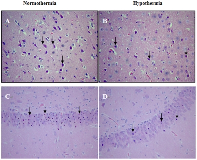
Figure 7. Representative microscopic changes in the cortex and hippocampus.
Hematoxylin and eosin stained sections (×200), in the precentral gyrus of the frontal lobe (A and B) and CA1 area of the hippocampus (C and D) at 24 h following ROSC, showed changes in damaged neurons, including an eosinophilic cytoplasm, loss of Nissl substance and nuclear pyknosis (arrows). The number of damaged neurons from hypothermic pigs (B and D) was markedly decreased when compared to normothermic pigs (A and C).

