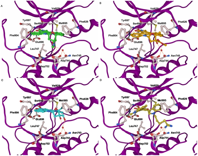
Figure 3. Details of the binding of (A) staurosporine (green), (B) K252a (orange), (C) lestaurtinib (cyan), and (D) Ro318220 (dark-yellow) to the PRK1 kinase domain.
Common interactions of the inhibitors are hydrogen bonds involving Glu696 and Ser698 of the hinge region, and van-der-Waals interactions with the gatekeeper residue Met695, as well as with Val629, Phe626, Leu747, and Phe904. In addition, some of the inhibitors interact with Asp744, Asn745 and a conserved water molecule (red sphere) nearby the Mg2+ binding site of the kinase. The backbone is shown as a purple ribbon. Only relevant amino acids are displayed. Hydrogen bonds are shown as dashed orange colored lines.

