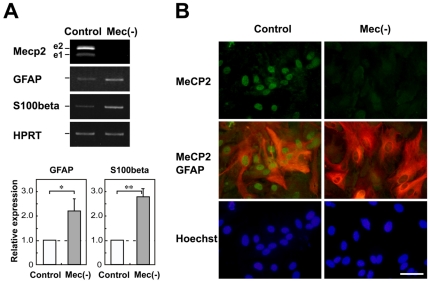
Figure 1. Characterization of assay cultures.
A. Expression of astroglial genes in primary cultured cortical astrocytes. Semi-quantitative RT-PCR analysis of Mecp2 and astroglial genes was performed in wild-type (white column) and MeCP2-null (gray column) astrocytes. Mecp2 e1 and e2 were detectable in the wild-type astrocytes. The lower graphs show that the GFAP/HPRT or S100β/HPRT expression ratio in each genotype was normalized against the level in control astrocytes. Bars represent the means ± standard errors (SE) of samples from three independent experiments (*p<0.05). The expression of astroglial markers was significantly upregulated by MeCP2 deficiency. B. Expression of MeCP2 in the primary cultured cortical astrocytes. The astrocytes were immunostained with MeCP2 (green) and GFAP (red) as glial-specific astrocytic markers. Scale bars indicate 50 µm.

