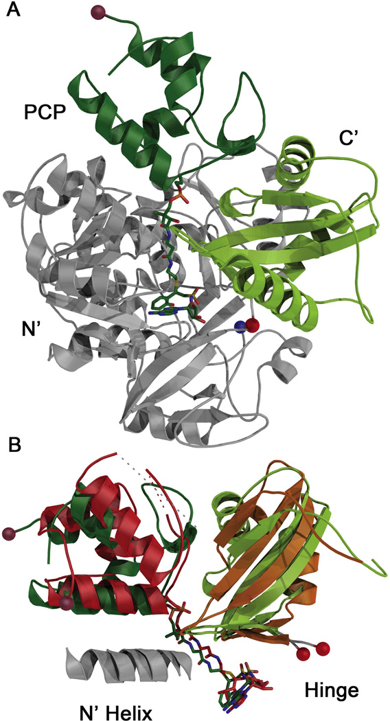
Figure 6.
Comparison of holo-PA1221 and EntE-B. (A) The interaction of the EntE-B protein (PDBID:3RG2) with N-terminal domain in grey, C-terminal subdomain is green, and PCP in forest green. The N- and C-termini, as well as the hinge residue, are shown with blue, brown, and red spheres respectively. The orientation is similar to the PA1221 orientation in Figure 3 (B) C-terminal subdomains and PCP domains of holo-PA1221 and EntE-B structurally aligned by least squares fitting of N-terminal domains, which are not shown for clarity. The N-terminal domain helices that interact with helix 2 of the PCP are shown in grey for both PA1221 and EntE-B and labeled N’ helix.

