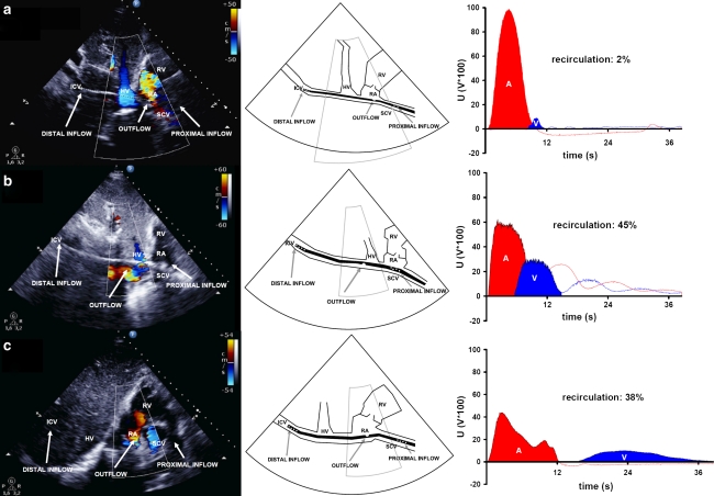
Fig. 1.
Transthoracic echocardiography (colour Doppler) shows: a the dual-lumen cannula (31 Fr.) situated properly in the caval veins (draining 4.3 L/min), with the outflow jet pointing at the tricuspid valve and right ventricle. The distance between the distal drainage port and the infusion port amounts to 9.4 cm (left). The schematic representation shows the position of the dual-lumen cannula (middle). Ultrasound dilution creates an arterial (A) and venous (V) dilution curve, resulting in a level of recirculation that amounts to 2 % (right). b An improperly situated dual-lumen cannula (27 Fr.). The cannula is rotated and the infusion port is located in the inferior caval vein near the hepatic vein (left). The schematic representation shows the position of the dual-lumen cannula (middle). Although ELS flow amounted to 4.7 L/min, ultrasound dilution measures a recirculation level of 45 % (right). c The bi-caval dual-lumen cannula (31 Fr.) correctly positioned. A large blue coloured jet within the superior caval vein is directed toward the proximal cannula drainage port and can be interpreted as drainage flow. A red coloured jet shows blood leaving the cannula infusion port and flowing directly toward the tricuspid valve (left). The schematic representation shows the position of the dual-lumen cannula (middle). Ultrasound dilution, however, detects a recirculation level of 38 % at a flow rate of 5.3 L/min, revealing cannula malposition (right). ICV inferior caval vein, SCV superior caval vein, RA right atrium, RV right ventricle, HV hepatic vein, A area under the curve measured by the arterial probe, V area under the curve measured by the venous probe, U voltages in volts × 100

