
Fig. 4.
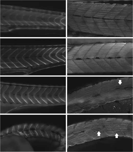
Myopathic features of megf10 morphant zebrafish muscle. Immunohistochemistry was performed with antibodies against β-dystroglycan (left) and myosin heavy chain (right) on wild-type fish and morphants treated with 6 ng COMO, 6 ng MO1, and 6 ng MO2 (top to bottom). Wild-type and COMO-treated fish showed myosepta with straight boundaries and tightly packed myofibers. Compared to control fish, MO1-treated fish showed subtle curving of myosepta and loosely packed myofibers with occasional prominent gaps (arrow), and MO2-treated fish showed less distinct somite boundaries and abundant gaps between myofibers (e.g., in somites indicated by arrows), creating an overall disorganization of the fiber pattern