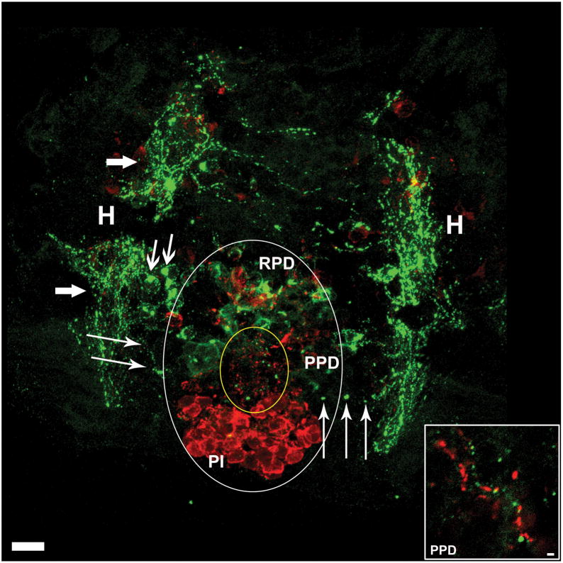
Figure 4. Hypothalamic AgRP and α-MSH Expressing Neurons Project to the Pituitary.
Horizontal view of larval zebrafish brain at 5dpf illustrating the pituitary and underlying hypothalamus. Large white oval outlines the extent of the pituitary, and small yellow oval indicates the anterior zone equivalent containing dense α-MSH immunoreactive (ir) fibers (red) and AgRP-ir fibers (green). Large arrows indicate hypothalamic AgRP-ir fiber bundles, medium arrows indicate hypothalamic AgRP-ir cell bodies, and thin arrows indicate AgRP-ir fibers projecting from hypothalamus into the pituitary. Inset is an enlargement from the PPD showing parallel AgRP-ir and α-MSH-ir neuronal fibers PI, pars intermedia; PPD, proximal pars distalis; RPD, rostral pars distalis; H, hypothalamus. Scale bars: main image =10μm, inset = 1μm.

