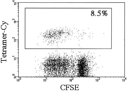
Figure 1.
Tetramer staining of human NK T cells. Human peripheral blood mononuclear cells were labeled with 5,6-carboxyfluorescein diacetate succinimidyl ester (CFSE) and cultured subsequently in RPMI medium 1640 supplemented with 10% (vol/vol) pooled human serum/100 units rIL-2/100 ng/ml αGalCer. After 4 days, cells were harvested and stained with a phycoerythrin-labeled Vβ11-specific antibody and a tricolor-labeled αGalCer–CD1 tetramer and analyzed on a FACScan (fluorescence-activated cell sorter). Both the Vβ11-specific antibody (data not shown) and the αGalCer–CD1 tetramer stained a similar population of cells, the majority of which had divided. The percentage of Vβ11/tetramer-positive cells at the start of the culture was 0.6%; after 7 days, it was 20% (data not shown).

