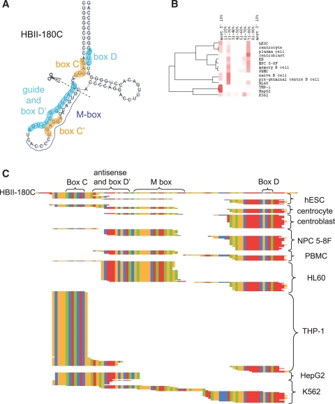
Figure 6.
Processing pattern of HBII-180C. (A) The predicted structure of HBII-180C is shown with boxes C and C′ highlighted in orange and boxes D′, D and guide regions highlighted in cyan. (B) Processing patterns of HBII-180C, derived as described in Figure 3. (C) Sequence alignment of HBII-180C and all its sdRNAs detected in cell types considered. Nucleotides are colour-coded: A (green), C (orange), G (red) and T (blue).

